
Substantia Nigra Pars Compacta
Question Dopamine Is Released Into The Striatum From The Substantia Nigra Pars Compacta Which Neural Response Would You Expect To See?.
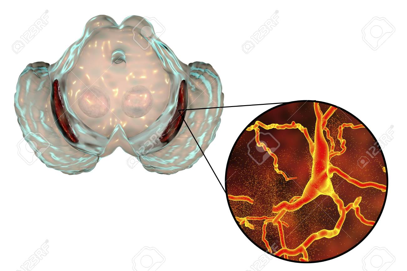
Substantia nigra pars compacta. The part of the substantia nigra that is heavily populated with pigmented dopamine neurons, which accounts for the darkened color of the substantia nigra when seen in unstained brain tissue The neurons of the pars compacta are more tightly packed together than those found in the pars reticulata (the other region of the substantia nigra) Learn more. Dopaminergic nerve cell bodies in such areas as the substantia nigra pars compacta tend to be pigmented due to the presence of the black pigment melanin, a direct chemical precursor to dopamine Dopaminergic pathways are involved in many functions such as executive function, learning, reward, motivation, and neuroendocrine control 3. The substantia nigra is a large pigmented cluster of neurons that consists of two parts, the pars reticulata and the pars compacta Cells of the pars compacta contain the dark pigment melanin;.
The part of the substantia nigra that is heavily populated with pigmented dopamine neurons, which accounts for the darkened color of the substantia nigra when seen in unstained brain tissue The neurons of the pars compacta are more tightly packed together than those found in the pars reticulata (the other region of the substantia nigra) Learn more. The mean retention latency for these groups ranged between sec In contrast, substantia nigra, pars compacta (SNC) ESB animals showed obvious disruption with a mean retention latency of 422 sec In another experiment post trial stimulation was equally effective. Left substantia nigra pars compacta activation (−4, −12, −8) greater in the time reproduction tasks (SHORT ± LONG) than the control reaction time task Activations are shown on the MNI reference brain, on sagittal, coronal, and horizontal views Parameter estimates for the left substantia nigra pars compacta showing increased activity.
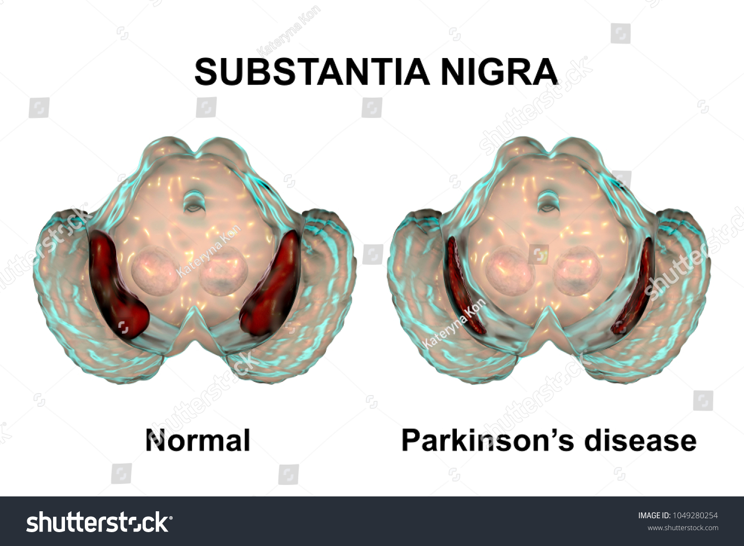
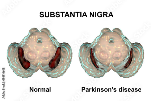
Substantia nigra in norm and in Parkinson's disease, 3D illustration showing decrease of its volume There is degeneration of dopaminergic neurons in the pars compacta of the substantia nigra Buy this stock illustration and explore similar illustrations at Adobe Stock. Dopaminergic nerve cell bodies in such areas as the substantia nigra pars compacta tend to be pigmented due to the presence of the black pigment melanin, a direct chemical precursor to dopamine Dopaminergic pathways are involved in many functions such as executive function, learning, reward, motivation, and neuroendocrine control 3. Pathophysiological dopamine depletion in the basal ganglia due to the death of dopaminergic (DA) neurons in the substantia nigra pars compacta (SNc) is related to the motor symptoms of PD (5, 6).
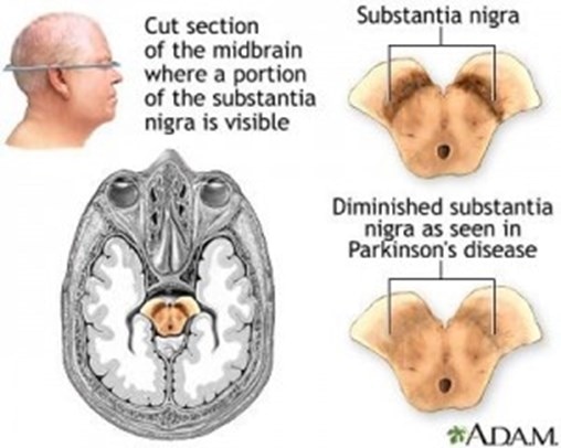
Communication among neurons of the substantia nigra pars compacta and the basal ganglia produce smooth, purposeful movement When the neurons in the substantia nigra are damaged in large numbers, the loss of dopamine prevents normal function in basal ganglia and causes the motor symptoms of PD tremor , rigidity , impaired balance , and loss of. Caused by a relatively selective loss of dopamine neurons in the substantia nigra pars compacta region of the midbrain These neurons are important for regulating motor function Normal Parkinson’s Disease Queens Square London Brain Bank Parkinson’s Disease (PD). 黒質緻密部 解剖学 黒質緻密部 (こくしつちみつぶ substantia nigra pars compacta)は、ヒトにおいて、ニューロメラニン色素を含有するニューロンが多く存在しているため黒色を帯びているが、加齢と共にニューロメラニンの量が減少する。 ニューロメラニンはドーパ(ヒドロキシフェニルアラニン.
Caused by a relatively selective loss of dopamine neurons in the substantia nigra pars compacta region of the midbrain These neurons are important for regulating motor function Normal Parkinson’s Disease Queens Square London Brain Bank Parkinson’s Disease (PD). Communication among neurons of the substantia nigra pars compacta and the basal ganglia produce smooth, purposeful movement When the neurons in the substantia nigra are damaged in large numbers, the loss of dopamine prevents normal function in basal ganglia and causes the motor symptoms of PD tremor , rigidity , impaired balance , and loss of. The properties of the hyperpolarizationactivated cation current (Ih) were investigated in rat substantia nigra pars compacta (SNc) principal neurons using patchclamp recordings in thin slices A reliable identification of single dopaminergic neurons was made possible by the use of a transgenic line of mice expressing eGFP under the tyrosine hydroxylase promoter.
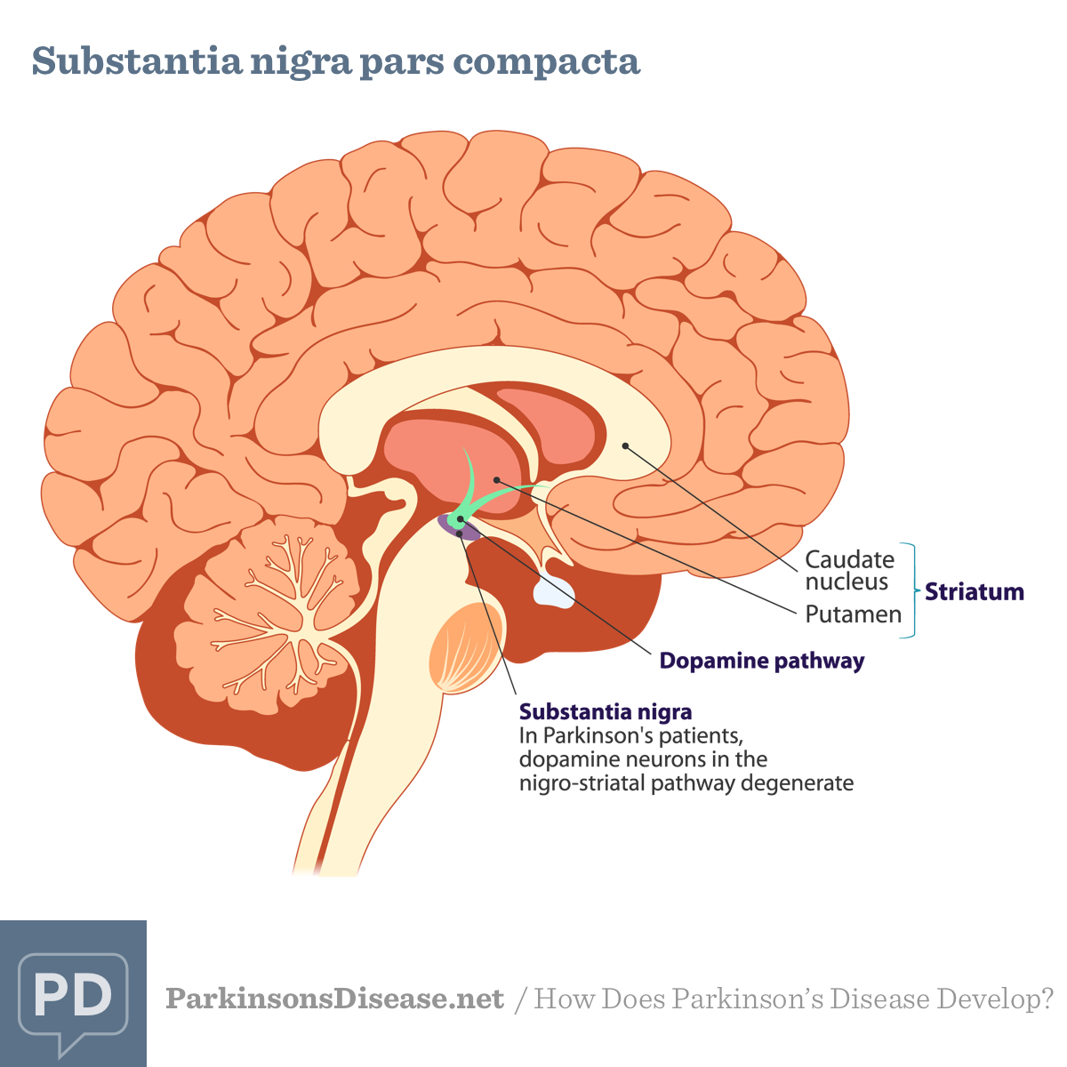
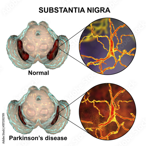
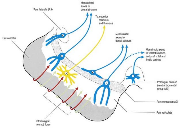
Most of the dopamine neurons of the brain originate in the midbrain and are found in either the substantia nigra or the ventral tegmental area, which is located adjacent to the substantia nigra The dopamine neurons in the substantia nigra express high levels of a pigment called neuromelanin, which accounts for their dark color These dopamine neurons, however, are found predominantly in the substantia nigra pars compacta The pars reticulata is instead populated largely by GABA neurons. Substantia nigra pars compacta (SNc) Neurons in the compacta are responsible for motor control Within this area are a large population of dopamine producing and releasing neurons These dopaminergic cells in the substantia nigra are often darker than the surrounding cells, which is how the region got named. In Parkinson’s disease, there is degeneration of neurons in the substantia nigra, mainly in pars compacta It results in decreased release of dopamine in the corpus striatum, leading towards the hypersensitivity of dopamine receptors in the striatum This disease is of unknown origin and affects the people between the ages of 45 and 55 years.
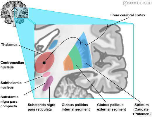
Caused by a relatively selective loss of dopamine neurons in the substantia nigra pars compacta region of the midbrain These neurons are important for regulating motor function Normal Parkinson’s Disease Queens Square London Brain Bank Parkinson’s Disease (PD). Left substantia nigra pars compacta activation (−4, −12, −8) greater in the time reproduction tasks (SHORT ± LONG) than the control reaction time task Activations are shown on the MNI reference brain, on sagittal, coronal, and horizontal views Parameter estimates for the left substantia nigra pars compacta showing increased activity. This pathway projects from the substantia nigra pars compacta to the striatum, and it utilizes the neurotransmitter dopamine This pathway has a modulatory effect on the basal ganglia, with dopamine facilitating the motor loop in these two ways It excites the direct pathway.
The substantia nigra, Latin for "black substance", or locus niger is a heterogeneous portion of the midbrain, separating the pes (foot) from the tegmentum (covering), and a major element of the basal ganglia system It consists of two strongly contrasted ensembles, the pars compacta and adjacent dopaminergic groups, and another ensemble made up of the pars reticulata and the pars lateralis. Caused by a relatively selective loss of dopamine neurons in the substantia nigra pars compacta region of the midbrain These neurons are important for regulating motor function Normal Parkinson’s Disease Queens Square London Brain Bank Parkinson’s Disease (PD). Activation Of The Frontal Cortex By Inhibiting The Direct Pathway And Inhibiting The Indirect Pathway Activation Of The Frontal Cortex By Activating The Direct Pathway And Inhibiting The Indirect Pathway Inhibition Of The Frontal.
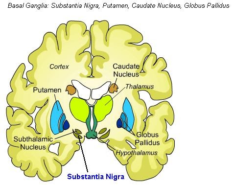
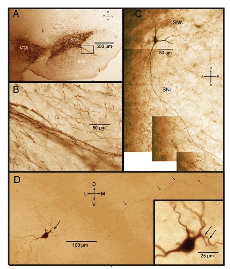
Dopamine neurons in the substantia nigra pars compacta (SNpc) were identified by antibodies to tyrosine hydroxylase (TH) and microglia were identified using Iba1 immunoreactivity The total number of TH neurons and the number of resting and activated microglia in the SNpc at 168 hours after the last dose were estimated using model or design. The substantia nigra is a distinct region within the basal ganglia, consisting of the substantia nigra pars compacta and the substantia nigra pars reticulata Both sections are characterized by a blackened appearance from a dark polymer pigment known as neuromelanin (Figs 1A and 1B) The dopamine transporter neurons reside in the substantia. Structures of the brain.
The part of the substantia nigra that is heavily populated with pigmented dopamine neurons, which accounts for the darkened color of the substantia nigra when seen in unstained brain tissue The neurons of the pars compacta are more tightly packed together than those found in the pars reticulata (the other region of the substantia nigra) Learn more. The substantia nigra (Latin for "black substance", Soemering) or locus niger is a heterogeneous portion of the midbrain, separating the pes (foot) from the tegmentum (covering), and a major element of the basal ganglia system It consists of two strongly contrasted ensembles, the pars compacta and adjacent dopaminergic groups, and another ensemble made up of the pars reticulata and the pars. The pars compacta is a portion of the substantia nigra, located in the midbrain It is formed by dopaminergic neurons and located medial to pars reticulata Parkinson's disease is characterized by the death of dopaminergic neurons in this region.
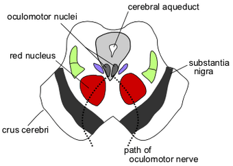
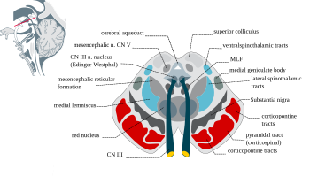
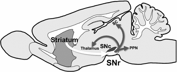
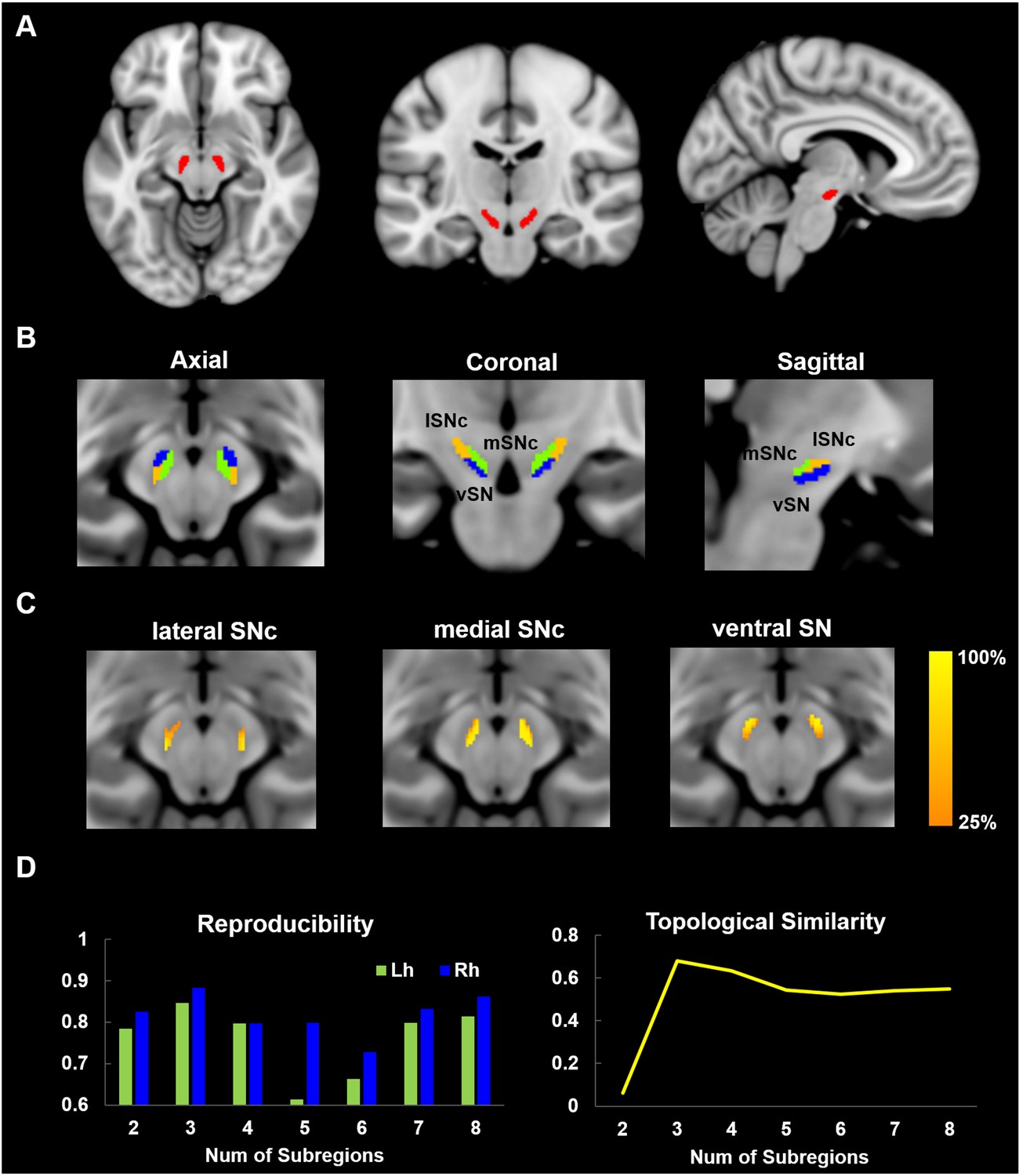
The substantia nigra is one of the brainstem nuclei and part of the extrapyramidal system While other nuclei such as the red nucleus are as small and contained within an axial slice at the superior colliculi (see figure), the substantia nigra is seen in axial slices at both superior and inferior colliculi. Introduction Parkinson disease (PD) is a common neurodegenerative disease characterized by disabling motor and nonmotor symptoms The pathologic correlate of nigrostriatal dopaminergic degeneration is the neuronal loss in substantia nigra (SN) pars compacta, particularly in the ventrolateral tier of neurons ()Standard magnetic resonance (MR) imaging techniques have a marginal role in the. The Substantia Nigra The substantia nigra (SN) is an area of deeply pigmented cells in the midbrain that regulates movement and coordination Neurons of the SN are divided into the substantia nigra pars compacta (SNc) and the substantia nigra pars reticulata (SNr).
Dopaminergic nerve cell bodies in such areas as the substantia nigra pars compacta tend to be pigmented due to the presence of the black pigment melanin, a direct chemical precursor to dopamine Dopaminergic pathways are involved in many functions such as executive function, learning, reward, motivation, and neuroendocrine control 3. Pathophysiological dopamine depletion in the basal ganglia due to the death of dopaminergic (DA) neurons in the substantia nigra pars compacta (SNc) is related to the motor symptoms of PD (5, 6). That is, they generate action potentials at a clocklike 2–4 Hz in the absence of synaptic input (Surmeier et al, 05) In this respect, they are much like cardiac pacemakers.
Substantia nigra pars compacta (SNc) dopamine neurons and their targets are involved in addiction and cueinduced relapse However, afferents onto SNc dopamine neurons themselves appear insensitive to drugs of abuse, such as cocaine, when afferents are collectively stimulated electrically This contrasts with ventral tegmental area (VTA) dopamine neurons, whose glutamate afferents react. Substantia nigra pars compacta (SNc) dopamine neurons and their targets are involved in addiction and cueinduced relapse However, afferents onto SNc dopamine neurons themselves appear insensitive to drugs of abuse, such as cocaine, when afferents are collectively stimulated electrically This contrasts with ventral tegmental area (VTA) dopamine neurons, whose glutamate afferents react. The part of the substantia nigra that is heavily populated with pigmented dopamine neurons, which accounts for the darkened color of the substantia nigra when seen in unstained brain tissue The neurons of the pars compacta are more tightly packed together than those found in the pars reticulata (the other region of the substantia nigra) Learn more.
The substantia nigra and its relations with the striatum in the monkey Progress in Brain Research 87 81–99 ↑ Hajos, M & Greenfield, SA (1994) Synaptic connections between pars compacta and pars reticulata neurones electrophysiological evidence for functional modules within the substantia nigra Brain Research 660 (2) 216–224. Dopaminergic nerve cell bodies in such areas as the substantia nigra pars compacta tend to be pigmented due to the presence of the black pigment melanin, a direct chemical precursor to dopamine Dopaminergic pathways are involved in many functions such as executive function, learning, reward, motivation, and neuroendocrine control 3. Caused by a relatively selective loss of dopamine neurons in the substantia nigra pars compacta region of the midbrain These neurons are important for regulating motor function Normal Parkinson’s Disease Queens Square London Brain Bank Parkinson’s Disease (PD).
The substantia nigra (SN) is an area of deeply pigmented cells in the midbrain that regulates movement and coordination Neurons of the SN are divided into the substantia nigra pars compacta (SNc) and the substantia nigra pars reticulata (SNr) Neurons of the SNc produce Dopamine, which stimulates movement. In all control subjects, the substantia nigra appeared as a hypointense area on the subtraction images, whose morphology closely matched known midbrain anatomy On these images, we were unable to distinguish the substantia nigra pars compacta and the pars reticulata 17 Figure 2 depicts 2 central sections from an MC and from a PD The outlines. The pars compacta forms one half of the substantia nigra, a region of the midbrain near the brain stem The primary function of the pars compacta is the production of a neurotransmitter called dopamineDopamine pertains to addiction, emotional responses and movement.
Substantia nigra The substantia nigra is a structure located in the mesencephalon (midbrain) that plays an important role in reward, addiction, and movement Substantia nigra is Latin for “black substance”, reflecting the fact that parts of the substantia nigra appear darker than neighboring areas due to high levels of neuromelanin in dopaminergic neurons. The part of the substantia nigra that is heavily populated with pigmented dopamine neurons, which accounts for the darkened color of the substantia nigra when seen in unstained brain tissue The neurons of the pars compacta are more tightly packed together than those found in the pars reticulata (the other region of the substantia nigra) Learn more. Background Up to the moment there is no universally accepted scheme of spatial organization of the groups of neurons of substantia nigra pars compacta of the human midbrain A detailed study of the architectonics of this structure is necessary for pathomorphological analysis of agerelated changes in the nervous tissue and the associated neurodegenerative diseases with selective death of.
黒質緻密部 解剖学 黒質緻密部 (こくしつちみつぶ substantia nigra pars compacta)は、ヒトにおいて、ニューロメラニン色素を含有するニューロンが多く存在しているため黒色を帯びているが、加齢と共にニューロメラニンの量が減少する。 ニューロメラニンはドーパ(ヒドロキシフェニルアラニン. A direct projection from superior colliculus to substantia nigra pars compacta in the cat Neuroscience 06;138(1) doi /jneuroscience Epub 05 Dec 19 Authors J G McHaffie 1 , H Jiang, P J May, V Coizet, P G Overton, B E Stein, P Redgrave Affiliation 1 Department of. The properties of the hyperpolarizationactivated cation current (Ih) were investigated in rat substantia nigra pars compacta (SNc) principal neurons using patchclamp recordings in thin slices A reliable identification of single dopaminergic neurons was made possible by the use of a transgenic line of mice expressing eGFP under the tyrosine hydroxylase promoter.
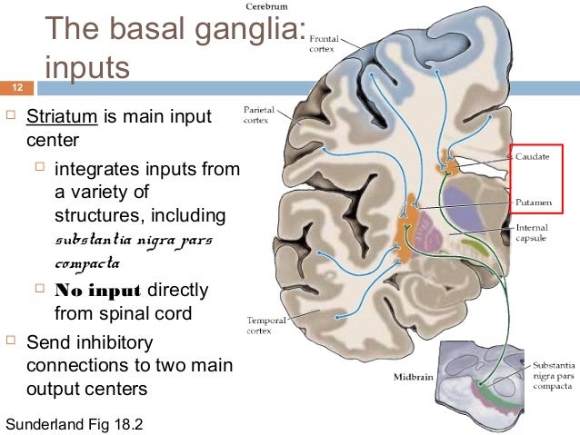
Activation Of The Frontal Cortex By Inhibiting The Direct Pathway And Inhibiting The Indirect Pathway Activation Of The Frontal Cortex By Activating The Direct Pathway And Inhibiting The Indirect Pathway Inhibition Of The Frontal. The dopaminergic tract is predominantly affected in Parkinson disease, and histologically, it is characterized by nigrostriatal dopaminergic degeneration leading to neuronal loss in the substantia nigra pars compacta, most conspicuous in the ventrolateral tier of neurons 11. These cells synthesize dopamine and project to either the caudate nucleus or the putamen By inhibiting the action of large aspiny striatal neurons in the caudate nucleus and the putamen (described above in.
Substantia substan´sheah (L) substance;. Pars compacta Substantia nigra pars compacta, utgör det dorsala skiktet av substantia nigra, är bland de mest undersökta cellgrupperna i de basala ganglierna och består av stora dopamininnehållande celler som känns igen på den mörka, neuromelanininducerade färgen. Above is part of a list of various areas and anatomical names of the human brain This anatomy is spelled and pronouncedTHANK YOU for WATCHING, SUBSCRIBING,.
Question Dopamine Is Released Into The Striatum From The Substantia Nigra Pars Compacta Which Neural Response Would You Expect To See?. The substantia nigra is divided into two parts the pars reticulata and pars compacta, which lies medial to the pars reticulata Sometimes a third region, the pars lateralis, is mentioned, though it is usually classified as part of the pars reticulata. The substantia nigra (Latin for "black substance", Soemering) or locus niger is a heterogeneous portion of the midbrain, separating the pes (foot) from the tegmentum (covering), and a major element of the basal ganglia system It consists of two strongly contrasted ensembles, the pars compacta and adjacent dopaminergic groups, and another ensemble made up of the pars reticulata and the pars.
Substantia nigra pars compacta (SNc) dopamine neurons are autonomously active;. Left substantia nigra pars compacta activation (−4, −12, −8) greater in the time reproduction tasks (SHORT ± LONG) than the control reaction time task Activations are shown on the MNI reference brain, on sagittal, coronal, and horizontal views Parameter estimates for the left substantia nigra pars compacta showing increased activity. The properties of the hyperpolarizationactivated cation current (I h) were investigated in rat substantia nigra pars compacta (SNc) principal neurons using patchclamp recordings in thin slices A reliable identification of single dopaminergic neurons was made possible by the use of a transgenic line of mice expressing eGFP under the tyrosine hydroxylase promoter.
Pars compacta Substantia nigra pars compacta, utgör det dorsala skiktet av substantia nigra, är bland de mest undersökta cellgrupperna i de basala ganglierna och består av stora dopamininnehållande celler som känns igen på den mörka, neuromelanininducerade färgen. These cells synthesize dopamine and project to either the caudate nucleus or the Read More;. Other articles where Pars compacta is discussed human nervous system Midbrain the pars reticulata and the pars compacta Cells of the pars compacta contain the dark pigment melanin;.
Used in anatomic nomenclature in naming various components of body tissues or structures substantia al´ba white matter substantia gelatino´sa the substance sheathing the posterior horn of the spinal cord and lining its central canal substantia gri´sea gray matter substantia ni´gra a dark layer of. The substantia nigra is a basal ganglia structure located in the midbrain that plays an important role in reward and movement Substantia nigra is Latin for "black substance", reflecting the fact that parts of the substantia nigra appear darker than neighboring areas due to high levels of neuromelanin in dopaminergic neurons Parkinson's disease is characterized by the loss of dopaminergic neurons in the substantia nigra pars compacta Although the substantia nigra appears as a continuous band i. The substantia nigra is one of the brainstem nuclei and part of the extrapyramidal system While other nuclei such as the red nucleus are as small and contained within an axial slice at the superior colliculi (see figure), the substantia nigra is seen in axial slices at both superior and inferior colliculi.
Substantia nigra pars compacta the part of the substantia nigra that is heavily populated with pigmented dopamine neurons, which accounts for the darkened color of the substantia nigra when seen in unstained brain tissue The neurons of the pars compacta are more tightly packed together than those found in the pars reticulata (the other region of the substantia nigra). DH Zald, in Brain Mapping, 15 Abstract Located in the midbrain, the substantia nigra (SN) can be divided into the pars compacta, which is the primary source of dopamine (DA) projections to the striatum, and the pars reticulata, which represents a key output zone, the basal ganglia The SN can be visualized in detail with specific MRI sequences and, with PET, radioligands targeting.
Pars Reticulata 978 613 3 9

The Autonomic Nervous System Ppt Download

Substantia Nigra Pars Compacta Anatomy Of The Brain Seehearsaylearn Youtube
Substantia Nigra Pars Compacta のギャラリー
Science Natural Phenomena Medicine Substantia Nigra

Know Your Brain Substantia Nigra Neuroscientifically Challenged

L 27 Basal Ganglia I Anatomy And Cell Physiology Flashcards Cram Com

Basal Ganglia Flashcards Quizlet

Article On Substantia Nigra Standard Of Care

Substantia Nigra Functions Location Stucture Clinical Significance

Corpus Striatum Neuroanatomy An Illustrated Colour Text 4 Ed
How Does Parkinson S Disease Develop
Ecurriculum Som Vcu Edu Portal Resources 09 Neuro Basalganglia Lecture Pdf

Substantia Nigra Wikipedia

Basal Ganglia
Substantia Nigra A Basal Banglia Of The Midbrain In Parkinson S Stock Photo Picture And Royalty Free Image Image
Three Dimensional Reconstruction Of Substantia Nigra Pars Compacta Of Human Brain Voronkov I P Pavlov Russian Medical Biological Herald
Q Tbn And9gcre1bbffxkkoczcf1uza928buceqdyd5zozsfz1tlsthn0w4gl7 Usqp Cau

Figure 1 The Involvement Of Neuroinflammation And Kynurenine Pathway In Parkinson S Disease

Substantia Nigra Wikiwand

Making Things Happen 2 Motor Disorders How Your Brain Works Week 7 Dr Jan Schnupp Howyourbrainworks Net Ppt Download
An Iron Dopamine Index Predicts Risk Of Parkinsonian Neurodegeneration In The Substantia Nigra Pars Compacta Chemical Science Rsc Publishing Doi 10 1039 C3sch
The Neurochemistry Of Parkinson S Disease Proteintech Group

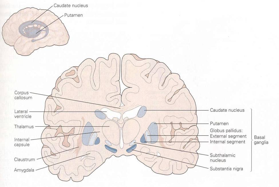
Figure 1 Coronal Section At The Level Parkinson S Disease Ncbi Bookshelf

Non Linear Developmental Trajectory Of Electrical Phenotype In Rat Substantia Nigra Pars Compacta Dopaminergic Neurons Elife

Nigrostriatal Pathway Wikipedia

Substantia Nigra Radiology Reference Article Radiopaedia Org

Parkinson S Disease Symptoms Causes Parkinsonism Mnc

Group Iii Metabotropic Glutamate Receptor Mediated Modulation Of Excitatory Transmission In Rodent Substantia Nigra Pars Compacta Dopamine Neurons Journal Of Pharmacology And Experimental Therapeutics

Neurodegeneration In The Substantia Nigra Pars Compacta Snpc Of Download Scientific Diagram

Jci Welcome

Dit Neuro 17 Flashcards Quizlet

2 Minute Neuroscience Substantia Nigra Youtube
Plos One Characterization Of Fetal Antigen 1 Delta Like 1 Homologue Expressing Cells In The Rat Nigrostriatal System Effects Of A Unilateral 6 Hydroxydopamine Lesion

Parkinson S Disease Anatomy Physiology

Ppt Basal Ganglia Powerpoint Presentation Free Download Id

An Iron Dopamine Index Predicts Risk Of Parkinsonian Neurodegeneration In The Substantia Nigra Pars Compacta Chemical Science Rsc Publishing

Substantia Nigra Wikipedia

Basal Ganglia

File Substantia Nigra Pars Compacta Jpg Wikimedia Commons

Aging Mildly Affects Dendritic Arborisation And Synaptic Protein Expression In Human Substantia Nigra Pars Compacta Sciencedirect
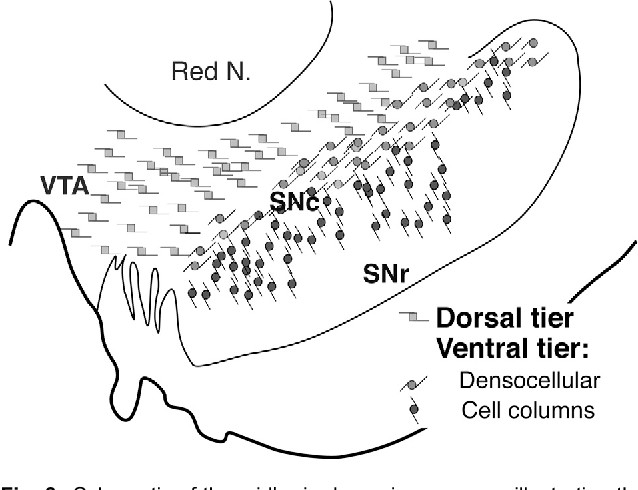
Figure 3 From The Place Of Dopamine In The Cortico Basal Ganglia Circuit Semantic Scholar
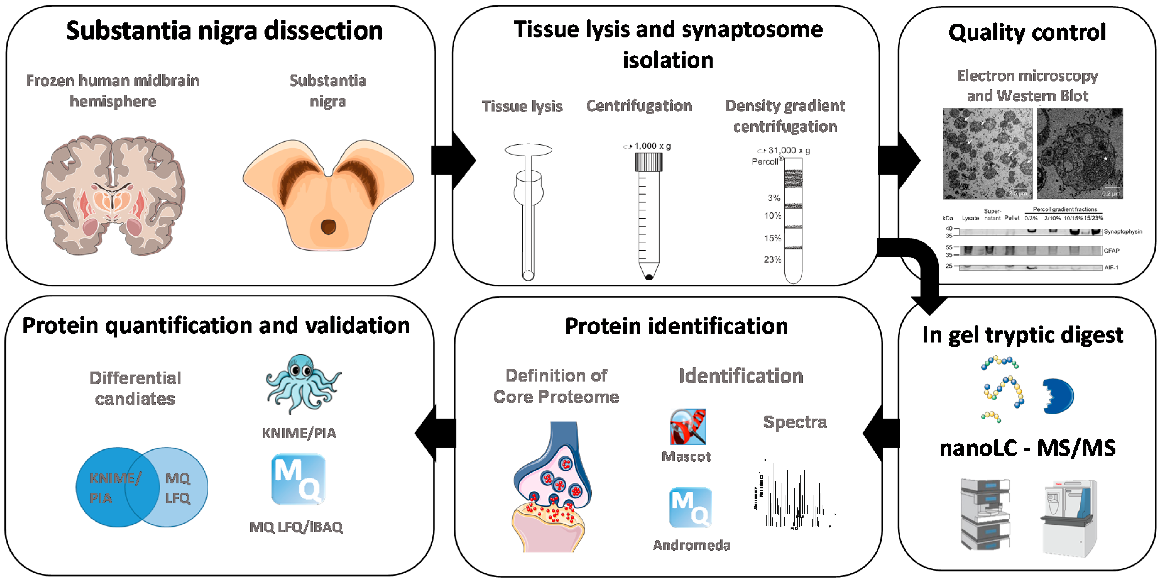
Cells Free Full Text Proteomic Characterization Of Synaptosomes From Human Substantia Nigra Indicates Altered Mitochondrial Translation In Parkinson S Disease
Basal Ganlia In A Neuroanatomy And A Neurophysiology Viewpoint

Substantia Nigra Pars Compacta An Overview Sciencedirect Topics
Plos One Noisy Galvanic Vestibular Stimulation Promotes Gaba Release In The Substantia Nigra And Improves Locomotion In Hemiparkinsonian Rats
Substantia Nigra Norm Parkinsons Disease 3d Stock Illustration

Substantia Nigra Functions Location Stucture Clinical Significance

Midbrain Iron Content In Early Parkinson Disease Neurology

Schematic Representation Of Different Sites In The Rat And Human Brains Download Scientific Diagram

Substantia Nigra Sheffield Neurogirls
Digitum Um Es Digitum Bitstream 101 133 1 Pathological changes in dendrites of substantia nigra neurons in parkinsons disease a golgi study Pdf
Substantia Nigra Psychology Wiki Fandom

Substantia Nigra In Parkinson S Disease 3d Illustration Showing Stock Photo Picture And Royalty Free Image Image

Pin On Person
Substantia Nigra
Substantia Nigra Control Of Basal Ganglia Nuclei Springerlink
Diseases Of Dopamine
Spontaneous Firing Properties Of Substantia Nigra Pars Compacta Snc Download Scientific Diagram

File Substantia Nigra Pars Compacta Pars Reticulata Png Wikimedia Commons
Substantia Nigra In Norm And In Parkinson S Disease 3d Illustration Showing Decrease Of Its Volume There Is Degeneration Of Dopaminergic Neurons In The Pars Compacta Of The Substantia Nigra Buy This

Effects Of Ketamine On Vocal Impairment Gait Changes And Anhedonia Induced By Bilateral 6 Ohda Infusion Into The Substantia Nigra Pars Compacta In Rats Therapeutic Implications For Parkinson S Disease Sciencedirect
Substantia Nigra Wikipedia

Immunohistochemical Analysis Of Th In The Pars Compacta Of Substantia Nigra Of Mice Injected With A Single I P Dose Of 30 Mg Kg Of Mptp Alone Or Combined With Ly 0 25 Or 3

Three Dimensional Reconstruction Of Substantia Nigra Pars Compacta Of Human Brain Voronkov I P Pavlov Russian Medical Biological Herald
Q Tbn And9gcsxkstqobxjisd2swv5bekpc3d95wz9suvizzxsdk4gzy0vnz6z Usqp Cau

Figure 1 Receptor Antagonism And Dyskinesia In Parkinson S Disease

Summary Of The Organization Of The Substantia Nigra Pars Reticulata And Download Scientific Diagram
Www Weizmann Ac Il Neurobiology Labs Ulanovsky Sites Neurobiology Labs Ulanovsky Files Uploads Intro Systemsneurosci Lecture13 Rivlin Basal Ganglia 30dec16 Pdf
Cerebellum And Basal Ganglia

What Is The Structure And Function Of The Substantia Nigra Brain Stuff
Substantia Nigra In Norm And In Parkinson S Disease 3d Illustration Showing Decrease Of Its Volume There Is Degeneration Of Dopaminergic Neurons In The Pars Compacta Of The Substantia Nigra Buy This
Substantia Nigra Wikipedia

Parkinson S Disease A Novel Mri Method For Determining Structural Changes In The Substantia Nigra Journal Of Neurology Neurosurgery Psychiatry

Simon Group
1

Snpc Substantia Nigra Pars Compacta
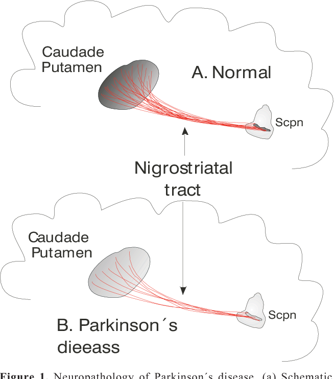
Figure 1 From Parkinson S Disease Molecular Aspects And Prospective Neuroprotective And Restorative Therapies Semantic Scholar
Participation Of The Pedunculopontine Tegmental Nucleus In Arousal Demanding Functions
Basal Ganglia Section 3 Chapter 4 Neuroscience Online An Electronic Textbook For The Neurosciences Department Of Neurobiology And Anatomy The University Of Texas Medical School At Houston

Bilateral And Partial Substantia Nigra Pars Compacta Snc Dopaminergic Download Scientific Diagram

Supplement To Brain Derived Neurotrophic Factor Is Required For The Establishment Of The Proper Number Of Dopaminergic Neurons In The Substantia Nigra Pars Compacta Journal Of Neuroscience

View Image
Http Www Bioline Org Br Pdf Ni

Snpc Substantia Nigra Pars Compacta By Acronymsandslang Com
What Does Vta Snc Mean Definition Of Vta Snc Vta Snc Stands For Ventral Tegmental Area Substantia Nigra Pars Compacta By Acronymsandslang Com

Retinoic Acid Counteracts Developmental Defects In The Substantia Nigra Caused By Pitx3 Deficiency Development

Function And Location Of Substantia Nigra A Diagrammatic Explanation Bodytomy
Q Tbn And9gctlr1 Slgiooeme2cytqzgs9u5p5nne7lpfm7cp7c4 Usqp Cau

Supplement To Brain Derived Neurotrophic Factor Is Required For The Establishment Of The Proper Number Of Dopaminergic Neurons In The Substantia Nigra Pars Compacta Journal Of Neuroscience

Red Lines Mark The Boundaries Enclosing The Substantia Nigra Pars Download Scientific Diagram
Basal Ganglia Neupsy Key
Neuronal Circuits And Physiological Roles Of The Basal Ganglia In Terms Of Transmitters Receptors And Related Disorders The Journal Of Physiological Sciences Full Text

44 Basicmedical Key

The Basal Ganglia Are Extensively Interconnected Skeletal Muscle
Anatomical And Functional Organization Of The Human Substantia Nigra And Its Connections Elife

View Image
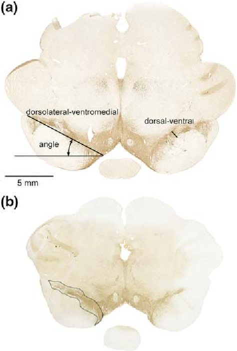
Figure 2 From The Substantia Nigra Pars Compacta Of The Gottingen Minipig An Anatomical And Stereological Study Semantic Scholar
Http Www Ajnr Org Content Early 10 12 23 Ajnr 355 Full Pdf
Plos One Transient Activation Of Gabab Receptors Suppresses Sk Channel Currents In Substantia Nigra Pars Compacta Dopaminergic Neurons

Figure 1 Electrical Synapses Between Dopaminergic Neurons Of The Substantia Nigra Pars Compacta Journal Of Neuroscience
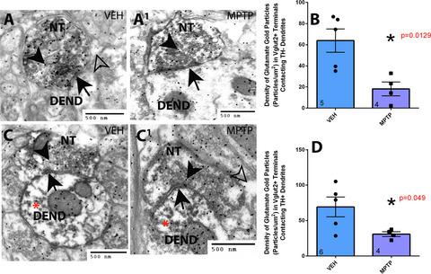
Differential Ultrastructural Alterations In The Vglut2 Glutamatergic Input To The Substantia Nigra Pars Compacta Pars Reticulata Following Nigrostriatal Dopamine Loss In A Progressive Mouse Model Of Parkinson S Disease European Journal Of Neuroscience
Plos One Assessment Of The Effects Of Mptp And Paraquat On Dopaminergic Neurons And Microglia In The Substantia Nigra Pars Compacta Of C57bl 6 Mice

Pin On Neuro 370
Brainpharm Neuron Substantia Nigra Pars Compacta Da Cell


