
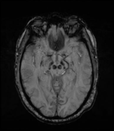
Substantia Nigra Mri
This study delineated longitudinal changes in different substantia nigra regions METHODS Seventytwo PD patients and 62 controls were studied at both baseline and after 18 months with MRI R2* and quantitative susceptibility mapping values from the substantia nigra pars compacta and substantia nigra pars reticulata were calculated.
Substantia nigra mri. Methods NMMRI and T 1weighted images were acquired from participants with cocaine use disorder and 35 control subjectsDiagnostic group effects in NMMRI signal were determined using a voxelwise analysis within the substantia nigra A subset of cocaine users and 17 control subjects also underwent functional MRI imaging using the monetary incentive delay task, in order to investigate. By depicting the white matter around the substantia nigra as an area of high signal intensity, diffusionweighted imaging shows the substantia nigra as a crescentshaped area of low signal intensity between the tegmentum of the midbrain and the cerebral peduncle. The MRI findings of substantia nigra edema in patients with SLE have not been previously reported Nonconvulsive status epilepticus can occur in patients with SLE and should be considered in.
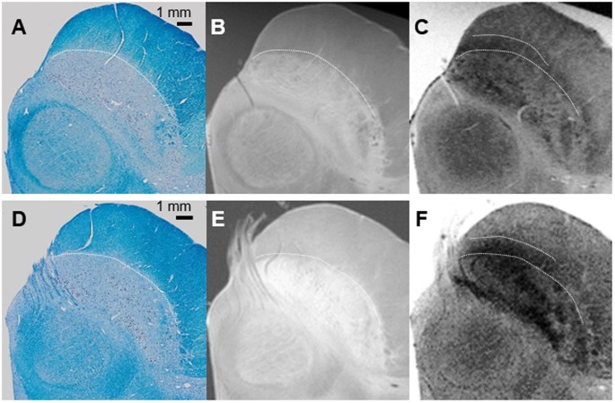
Article abstract—Elevated iron levels in the substantia nigra (SN) of the brain in Parkinson's disease (PD) may mediate lipid peroxidative reactions, promoting SN neuronal death To assess SN iron accumulation in living PD patients and its relation to motor performance, we measured, in 13 nondemented PD patients and 10 normal control subjects, simple reaction time (SRT) and simple movement. Lehericy S , Bardinet E , Poupon C , Vidailhet M , Francois C (14) 7 Tesla magnetic resonance imaging A closer look at substantia nigra anatomy in Parkinson’s disease Mov Disord 29, 1574–1581 33. 2) Due to its relatively small size, identifying SN in MR images is challenging The authors use a substantia nigra ROI obtained from a publicly available 7T MRI atlas and register this to every individual subject It would be good for the authors to illustrate (maybe in a figure supplement), how well this approach works.
Diffusion MRI may be able to help confirm tissue microstructural alterations in the substantia nigra to probe for the presence of asymmetry Purpose To investigate lateral asymmetry in the SN of patients with PD by using diffusion MRI with both Gaussian and nonGaussian models. Researchers have found that NMMRI signal is lower in the substantia nigra of people with Parkinson’s disease, reflecting the cell death that occurs in these patients. Recent Posts Happy New Year from Journey with Parkinson’s January 1, 21;.
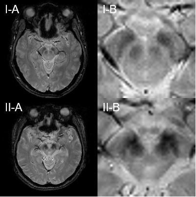
Conclusions Given that previous imaging studies show decreased dopamine signaling in the striatum, the finding of increased NMMRI signal in the substantia nigra provides additional insight into the pathophysiology of cocaine use disorder One interpretation is that cocaine use disorder is associated with a redistribution of dopamine between cytosolic and vesicular pools, leading to increased accumulation of neuromelanin. Differentiating earlystage Parkinson's disease (PD) from essential tremor (ET) remains challenging In the current study, we aimed to evaluate whether visual analyses of neuromelaninsensitive magnetic resonance imaging (NMMRI) combined with nigrosome1 (N1) imaging using quantitative susceptibility mapping (QSM) in the substantia nigra (SN) are of diagnostic value in the differentiation of. Additionally, the researchers validated a voxelwise NMMRI approach to determine substantia nigra subregions Using this approach along with molecular PET and functional MRI data, they further.
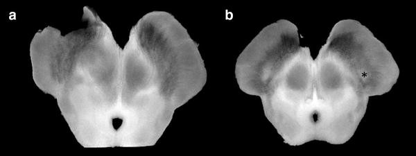
2) Due to its relatively small size, identifying SN in MR images is challenging The authors use a substantia nigra ROI obtained from a publicly available 7T MRI atlas and register this to every individual subject It would be good for the authors to illustrate (maybe in a figure supplement), how well this approach works. Diagnostic group effects in NMMRI signal were determined using a voxelwise analysis within the substantia nigra A subset of cocaine users and 17 control subjects also underwent functional MRI imaging using the monetary incentive delay task, in order to investigate whether NMMRI signal was associated with alterations in reward processing. The MRI results indicated there were higherthanusual iron deposits in the MPTPlesion side of the substantia nigra compared with the opposite side in the same animal Similar results were found when these animals were compared with the control group of monkeys, which had been injected with a saline solution.
Secondary degeneration of the substantia nigra and the corticospinal tract is demonstrated as a hyperintensity lesion on diffusionweighted imaging (DWI) during the subacute phase of ipsilateral striatal infarction (13) Four patients with typical DWI findings, including two who underwent followup magnetic resonance imaging (MRI) in the chronic. A specific T1weighted magnetic resonance imaging (MRI) sequence has been shown to detect substantia nigra (SN) neuromelanin (NM) signal changes that accurately discriminate Parkinson’s disease (PD) patients from controls, even in early disease stages. Huber SJ, Chakeres DW Magnetic resonance imaging in Parkinson's disease Arch Neurol 1990; 23 Pujol J, Junque C, Vendrell P, Grau JM, Capdevila A Reduction of the substantia nigra width and motor decline in aging and Parkinson's disease Arch Neurol 1992; 24 Stern MB, Braffman BH, Skolnick BE, Hurtig HI, Grossman RI.
This is where it gets its name, which is Latin for "black substance" Although it is often referred to as one structure,. Botulinum Toxin for Treatment of Dystonia. This atlas takes advantage of ultrahigh resolution 7T MRI to provide unprecedented levels of detail on structures of the basal ganglia invivo The ATAG atlas includes probability maps of the striatum, GPe, GPi, red nucleus, substantia nigra, subthalamic Nucleus(STh) and the PAG.
Substantia nigra R2* was higher at 1 year if ipsilateral striatum was infarcted than if not infarcted (P < 001) R2* mapping may be added to the followup MRI after stroke to monitor neurodegeneration by assessing iron deposition in the substantia nigra. This is where it gets its name, which is Latin for "black substance" Although it is often referred to as one structure,. The MRI results indicated there were higherthanusual iron deposits in the MPTPlesion side of the substantia nigra compared with the opposite side in the same animal Similar results were found when these animals were compared with the control group of monkeys, which had been injected with a saline solution.
Objective To detect changes in the different subregions of the substantia nigra pars compacta (SNc) in patients with Parkinson’s disease (PD) and to correlate the results with clinical data Background Reduced midbrain Neuromelanin (NM) in PD is attributed to loss of pigmented dopaminergic neurons localized especially to the SNc Noninvasive Magnetic Resonance Imaging (MRI) markers. To evaluate the anatomy of the substantia nigra (SN) in healthy subjects by performing 7T magnetic resonance (MR) imaging of the SN, and to prospectively define the accuracy of 7T MR imaging in distinguishing Parkinson disease (PD) patients from healthy subjects on an individual basis Materials and Methods. Abstract This study aimed to investigate the spatiotemporal changes in neuromelaninsensitive MRI signal in the substantia nigra and their relation to clinical scores of disease severity in patients with early or progressing Parkinson’s disease and patients with idiopathic rapid eye movement sleep behaviour disorder (iRBD) exempt of Parkinsonian signs compared to healthy control subjects.
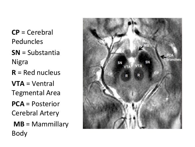
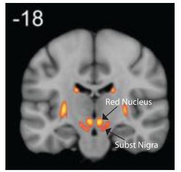
Excessive midbrain iron accumulation in Parkinson’s Disease (PD) contributes to degeneration of the substantia nigra pars compacta (SNc) and ventral tegmental area (VTA) Despite this understanding, there are no validated PD biomarkers Magnetic resonance imaging (MRI) can localize and quantify brain iron for diagnosis of PD Seventeen earlystage PD patients and twentyone controls were. The substantia nigra is one of the brainstem nuclei and part of the extrapyramidal system While other nuclei such as the red nucleus are as small and contained within an axial slice at the superior colliculi (see figure), the substantia nigra is seen in axial slices at both superior and inferior colliculi. Substantia nigra pars compacta (SNpc) and locus coeruleus (LC) are key brain areas involved in Parkinson’s disease The loss of important chemicals (dopamine and norepinephrine) made by cells in these areas leads to many of the symptoms of Parkinson’s disease.
The substantia nigra (“black substance” in Latin) is a long nucleus located in the midbrain but considered functionally a part of the basal ganglia because of its reciprocal connections with other brainstem nuclei It consists of two components, the pars compacta and the pars reticulata, which have different connections and use different neurotransmitters. BACKGROUND AND PURPOSE Visualizing with MR imaging and obtaining quantitative indexes of degeneration of the substantia nigra in Parkinson disease have been longsought goals We investigated the potential role of area and T1 contrast measurements in differentiating patients from controls and their agerelated changes. Neuromelaninsensitive MRI (NMMRI) purports to detect the content of neuromelanin (NM), a product of dopamine metabolism that accumulates in the substantia nigra (SN) Prior work has shown that NMMRI provides a marker of SN integrity in Parkinson’s disease Here, we show that it may additionally provide a marker of dopamine function in the human nigrostriatal pathway.
Substantia nigra and Parkinson disease Overview Parkinson disease is a slowly progressive disorder that affects movement, muscle control, and balance Part of the disease process develops as cells are destroyed in certain parts of the brain stem, particularly the crescentshaped cell mass known as the substantia nigra. Article abstract A 33yearold woman admitted for meningoencephalitis had features of encephalitis lethargica develop on her third day of illness She had ophthalmoplegia, akinetic mutism, and prominent extrapyramidal signs consisting of lip and hand tremors, cogwheel rigidity, and facial bradykinesia. Neuromelaninsensitive MRI (NMMRI) purports to detect the content of neuromelanin (NM), a product of dopamine metabolism that accumulates in the substantia nigra (SN) Prior work has shown that NMMRI provides a marker of SN integrity in Parkinson’s disease Here, we show that it may additionally provide a marker of dopamine function in the human nigrostriatal pathway.
Correia Guedes L , Reimão S , Paulino P , Nunes RG , BouçaMachado R , Abreu D , et al Neuromelanin magnetic resonance imaging of the substantia nigra in LRRK2 related Parkinson’s disease Mov Disord 17;–3 doi /mds270 15 Xing Y , Sapuan A , Dineen RA , Auer DP. The researchers also found that NMMRI signal in the substantia nigra was associated with functional MRI measures of regional cerebral blood flow Lastly, the researchers examined the link between. In Parkinson’s disease, the depletion of ironrich dopaminergic neurons in substantia nigra ’s nigrosome 1 precedes first motor symptoms by two decades Monitoring this neuronal depletion at an early disease stage is needed for diagnosis and treatment monitoring Magnetic resonance imaging (MRI) is particularly suitable for this task due to its sensitivity to tissue iron.
Parkinson’s disease (PD) is a progressive neurodegenerative disorder in which the major pathologic substrate is a loss of dopaminergic neurons from the substantia nigra Our main objective was to determine the correspondence between changes in the substantia nigra, evident in neuromelanin and iron sensitive magnetic resonance imaging (MRI), and dopaminergic striatal innervation loss in. Wellness Checklist for Life in the Presence of Parkinson’s December 27, ;. An ideal imaging marker would directly mirror the loss of substantia nigra dopaminergic neurones, which is most prominent in subregions called nigrosomes Highresolution, ironsensitive, magnetic resonance imaging (MRI) at 7T allows direct nigrosome1 visualisation in healthy people but not in PD.
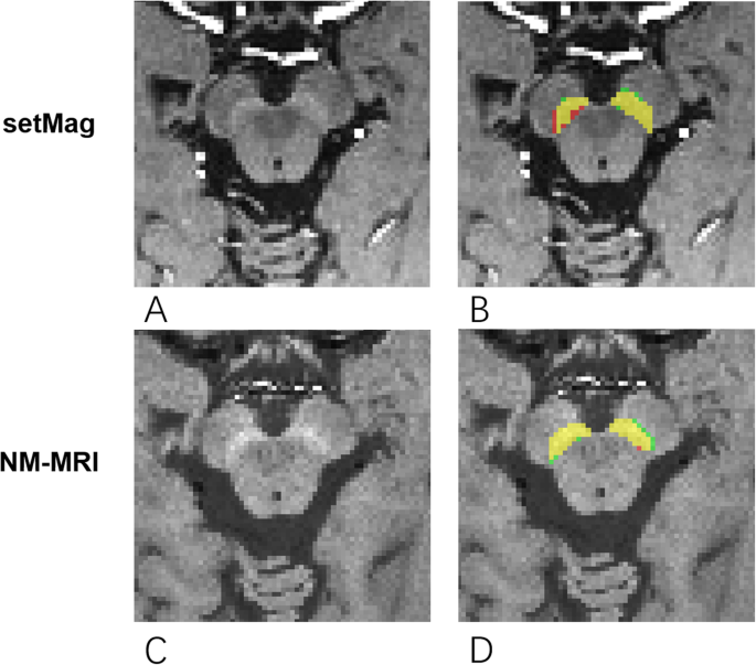
Neuromelanin‐sensitive MRI (NM‐MRI) of the substantia nigra provides a noninvasive way to acquire an indirect measure of dopamine functioning Despite the potential of NM‐MRI as a candidate biomarker for dopaminergic pathology, studies about its reproducibility are sparse. Brief Report Social Isolation and Loneliness are Risk Factors for Worsening Parkinson’s Symptoms December 28, ;. Study RationaleSubstantia nigra pars compacta (SNpc) and locus coeruleus (LC) are key brain areas involved in Parkinson’s disease The loss of important chemicals (dopamine and norepinephrine) made by cells in these areas leads to many of the symptoms of Parkinson’s disease Our group develops and uses magnetic resonance imaging (MRI) techniques that can measure changes in these brain areas.
To enhance the visibility of nigrosome 1 in substantia nigra, which has recently been suggested as an imaging biomarker for Parkinson's disease (PD) at 3T magnetic resonance imaging (MRI) Materials and Methods The substantia nigra structure was visualized at 3T MRI using multiecho susceptibility map‐weighted imaging (SMWI) in 15 healthy volunteers and 6 patients with Parkinson's disease (PD). The substantia nigra is a region in the midbrain that is considered part of the basal ganglia It looks like a darkened streak in unstained brain tissue;. Background Iron is important in the pathophysiology of Parkinson’s disease (PD) specifically related to degeneration of the substantia nigra (SN) Magnetic resonance imaging (MRI) can be used to measure brain iron in the entire structure but this approach is insensitive to regional changes in iron contentObjective The goal of this work was to use quantitative susceptibility mapping (QSM) and R2∗ to quantify both global and regional brain iron in PD patients and healthy controls (HC.
Thus, substantia nigra (SN) FA and R2* measures may reflect different aspects of PDrelated pathology, each with clinical implications Discovery Demonstrate that nigrostriatal DTI and R2* differentiate PD from parkinsonian syndromes Many conditions can disrupt basal ganglia function and mimic PD clinically eg progressive supranuclear palsy. Functional MRI (fMRI) has revealed that unpredictability of task order and random timing activate substantia nigra (Dreher and Grafman, 02), and neurons in the SNc show a depression in firing when an expected and timepredicted reward is not delivered (Hollerman and Schultz, 1998). Specifically, the NMMRI CNR in substantia nigra correlated positively with raclopride displacement in the dorsal striatum from the D 2 receptor by endogenous dopamine released by amphetamine, which acts on presynaptic vesicular and cytosolic pools in dopamine neurons This suggests that individual differences in the size of these releasable.
Kim JM, Jeong HJ, Bae YJ, et al Loss of substantia nigra hyperintensity on 7 Tesla MRI of Parkinson’s disease, multiple system atrophy, and progressive supranuclear palsy Parkinsonism Relat Disord, 16,2647–54 CrossRef Google Scholar. The combination of substantia nigra volume, signal intensity, and fractional anisotropy in the neuromelaninsensitive substantia nigra allowed excellent diagnostic accuracy (093) Visual assessment of both substantia nigra dorsolateral hyperintensity and neuromelaninsensitive images had good diagnostic accuracy (091 and 086, respectively). The substantia nigra is a basal ganglia structure located in the midbrain that plays an important role in reward and movement Substantia nigra is Latin for "black substance", reflecting the fact that parts of the substantia nigra appear darker than neighboring areas due to high levels of neuromelanin in dopaminergic neurons Parkinson's disease is characterized by the loss of dopaminergic neurons in the substantia nigra pars compacta Although the substantia nigra appears as a continuous band i.
Neuromelaninsensitive imaging T 1weighted MRI modality that is sensitive to neuromelanincontaining dopaminergic neurons in the substantia nigra which bind iron, forming paramagnetic neuromelaniniron complexes. Quotes to Help the Life Journey With Parkinson’s December 29, ;. Article abstract A 33yearold woman admitted for meningoencephalitis had features of encephalitis lethargica develop on her third day of illness She had ophthalmoplegia, akinetic mutism, and prominent extrapyramidal signs consisting of lip and hand tremors, cogwheel rigidity, and facial bradykinesia.
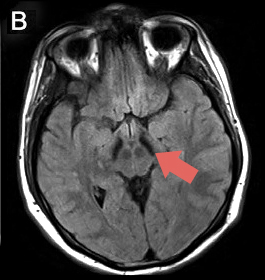
Neuromelanin‐sensitive MRI (NM‐MRI) of the substantia nigra provides a noninvasive way to acquire an indirect measure of dopamine functioning Despite the potential of NM‐MRI as a candidate biomarker for dopaminergic pathology, studies about its reproducibility are sparse Purpose. Our findings suggest that the CEST MRI signal of the substantia nigra is a potential imaging biomarker for the diagnosis and monitoring of the severity of PD Introduction Parkinson's disease (PD) is a common progressive neurodegenerative disease characterized by the loss of dopaminergic neurons in the substantia nigra ( Fearnley and Lees, 1991 ). Substantia Nigra Swelling and Dentate Nucleus T2 Hyperintensity May Be Early Magnetic Resonance Imaging Signs of β‐Propeller Protein‐Associated Neurodegeneration Camilla Russo MD Department of Advanced Biomedical Sciences, “Federico II” University of Naples, Naples, Italy.
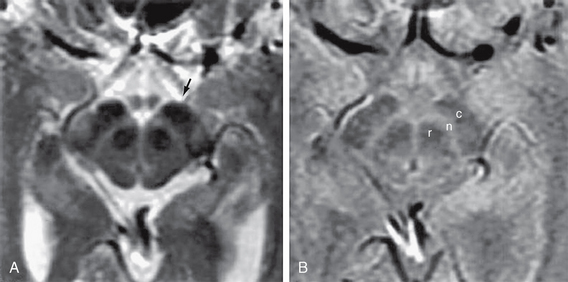
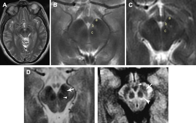
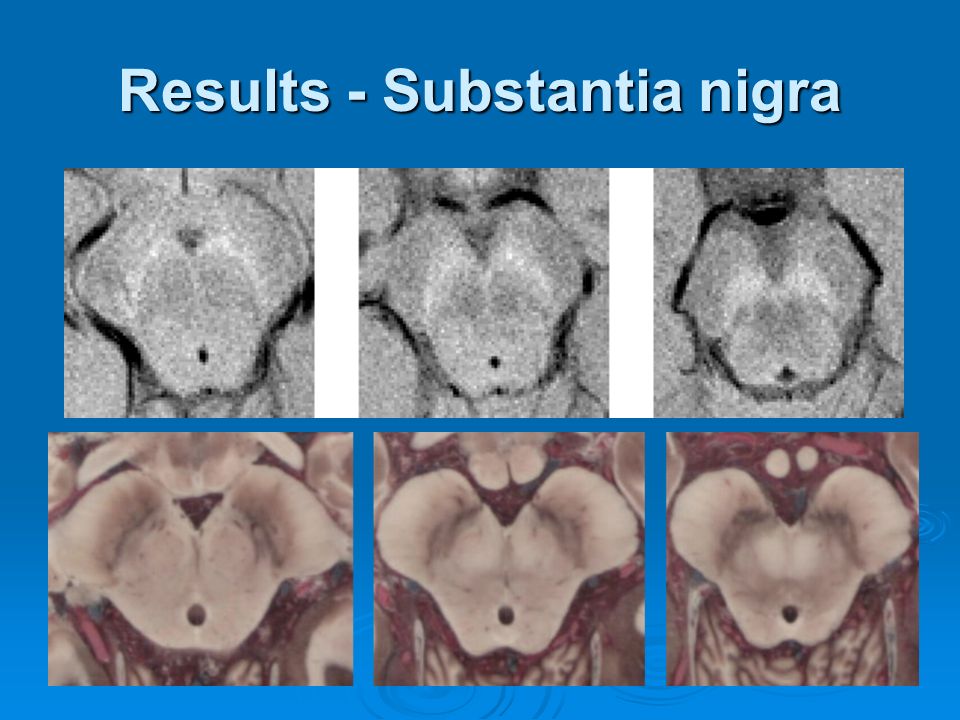
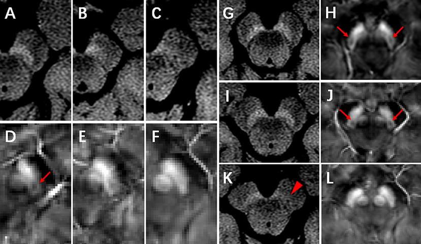
The swallow tail sign describes the normal axial imaging appearance of nigrosome1 within the substantia nigra on highresolution T2*/SWI weighted MRI Terminology Yes, this is one of those annoying signs where its presence is normal A normal. An ideal imaging marker would directly mirror the loss of substantia nigra dopaminergic neurones, which is most prominent in subregions called nigrosomes Highresolution, ironsensitive, magnetic resonance imaging (MRI) at 7T allows direct nigrosome1 visualisation in healthy people but not in PD. Editor In the June 14 issue of Radiology, Dr Cosottini and colleagues ( 1) describe altered appearances of the substantia nigra (SN) on susceptibilityweighted 7T magnetic resonance (MR) images in patients with Parkinson disease (PD) A threetier SN organization with a central highsignalintensity tier outlined by lowsignalintensity tiers was seen in healthy control subjects but not in patients with PD, enabling excellent discrimination.
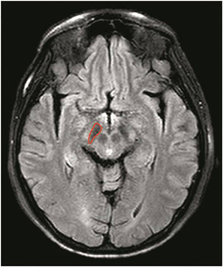
Coregistration of highresolution images with a digitized anatomic atlas revealed localization of secondary lesions primarily in the substantia nigra pars compacta Apparent diffusion coefficient (ADC) values in the secondary lesions showed a delayed sharp decline through day 10 Normalization of ADC values was observed at late measurements. The substantia nigra is a region in the midbrain that is considered part of the basal ganglia It looks like a darkened streak in unstained brain tissue;. Additionally, the researchers validated a voxelwise NMMRI approach to determine substantia nigra subregions Using this approach along with molecular PET and functional MRI data, they further.
Anatomical And Functional Organization Of The Human Substantia Nigra And Its Connections Elife

Substantia Nigra Scans 2 Of 2 Image Eurekalert Science News
Q Tbn And9gcrzg L7h7lroe3qdghdohf1bae Frdcb4nuojm8hpcwoh9ide Usqp Cau
Substantia Nigra Mri のギャラリー

Simultaneous Imaging Of Locus Coeruleus And Substantia Nigra With A Quantitative Neuromelanin Mri Approach Sciencedirect

Effects Of Parkinson S Disease Mutation Reversed In Cells Neuroscience News
Reproducibility Of Locus Coeruleus And Substantia Nigra Imaging With Neuromelanin Sensitive Mri Springerlink
Mri Findings Nbia
Www Strokejournal Org Article S1052 3057 15 0 Pdf
Neurodegeneration Cerebellum And Brain Stem Radiology Key

Neuromelanin Sensitive Mri Identified As A Potential Biomarker For Psychosis

In This Figure We Have Displayed Two Images Containing The Substantia Download Scientific Diagram
Q Tbn And9gcsrlizvfwvuo7dkfggxoxzpayrrwu5skf6 Er2cbokiseh6imww Usqp Cau

Magnetic Resonance Imaging T2 Weighted Axial Images Sh Open I

T2 Weighted Axial Magnetic Resonance Imaging Showing Bilateral Isolated Download Scientific Diagram

Changes In Neuromelanin Mri Signal In Parkinson S Disease A Longitudinal Study Mds Abstracts

Substantia Nigra Wikipedia
Plos One Dopaminergic Neurodegeneration In The Mouse Is Associated With Decrease Of Viscoelasticity Of Substantia Nigra Tissue
Ismrm Digital Posters Page Neuro Neurodegeneration

The Role Of Diffusion Magnetic Resonance Imaging In Parkinson S Disease And In The Differential Diagnosis With Atypical Parkinsonism


Pdf Identifying The Functional Architecture Of The Human Ventral Tegmental Area And The Substantia Nigra Using High Resolution Magnetic Resonance Imaging Semantic Scholar
Case Report
Mri Findings Nbia
English T2 Weighted Mri Sequences Demonstrating Globus Pallidus Hypointensity A And Hypointensity Of The Substantia Nigra B Arrows In Bpan The Substantia Nigra Is Usually More Hypointense Relative To The Globus Pallidus Indicating
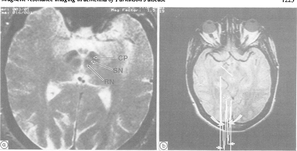
Figure 2 From Dementia Of Parkinson S Disease Magnetic Resonance Imaging In Semantic Scholar
Http Portal Research Lu Se Portal Files 1 G5 Yulia S Pdf

The Role Of Diffusion Magnetic Resonance Imaging In Parkinson S Disease And In The Differential Diagnosis With Atypical Parkinsonism

Parkinsonism Due To Predominant Involvement Of Substantia Nigra In Japanese Encephalitis Neurology

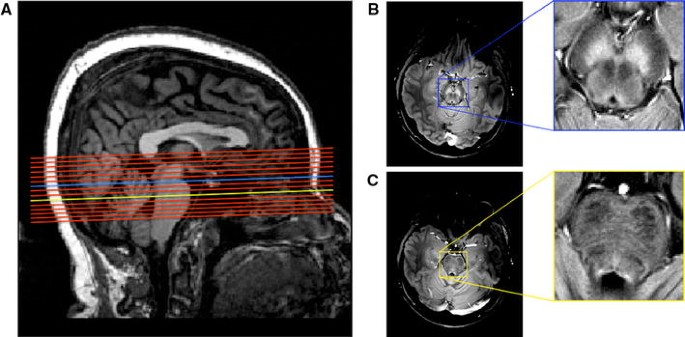
Ppt Mri Of The Locus Coeruleus And Substantia Nigra Powerpoint Presentation Id
Imaging Parkinsonian Pathology In Substantia Nigra With Mri Springerlink

Secondary Change In The Substantia Nigra Induced By Incomplete Infarct And Minor Hemorrhage In The Basal Ganglia Due To Traumatic Middle Cerebral Arterial Dissection Stroke
Http Www Bioline Org Br Pdf Ni
Figure 1 From T2 Weighted Mri In Parkinson S Disease Substantia Nigra Pars Compacta Hypointensity Correlates With The Clinical Scores Semantic Scholar
Anatomy Of The Substantia Nigra And Subthalamic Nucleus On Mr Imaging Radiology Key

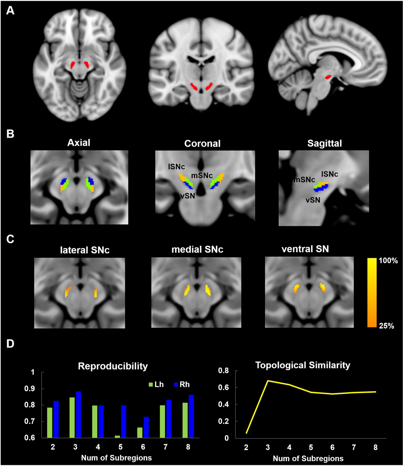
Parcellation Of The Human Substantia Nigra Based On Anatomical Connectivity To The Striatum Sciencedirect

View Image

Mr Imaging Of The Substantia Nigra At 7 T Enables Diagnosis Of Parkinson Disease Radiology
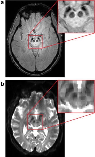
Short Echo Time Magnitude Image Derived From Quantitative Susceptibility Mapping Could Resemble Neuromelanin Sensitive Mri Image In Substantia Nigra Bmc Neurology Full Text
Nanopdf Com Download A Axial T2 Weighted Mri Scan Through The Lateral Ventricles Of Abnormal High Sig Pdf
Presentation1 Pptx Radiological Imaging Of Parkinsonism
Core Ac Uk Download Pdf Pdf

Axial Mri Substantia Nigra Diagram Quizlet
Mri Findings Nbia
Parkinson Disease Radiology Reference Article Radiopaedia Org
The Neuromelanin Related T 2 Contrast In Postmortem Human Substantia Nigra With 7t Mri Scientific Reports

Parkinson S Disease A Novel Mri Method For Determining Structural Changes In The Substantia Nigra Journal Of Neurology Neurosurgery Psychiatry

The Substantia Nigra Pars Compacta And Temporal Processing Journal Of Neuroscience

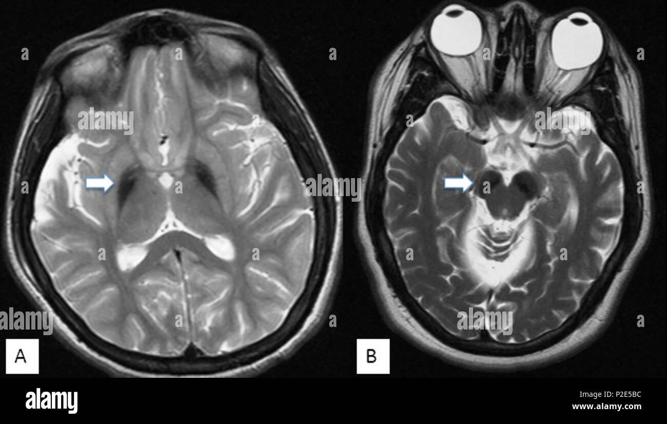
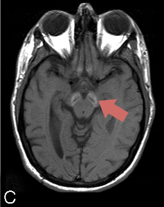
Bilateral Substantia Nigra Lesions On Magnetic Resonance Imaging In A Patient With Encephalitis Lethargica Journal Of Neurology Neurosurgery Psychiatry

Figure 1 From Chemical Exchange Saturation Transfer Mri Signal Loss Of The Substantia Nigra As An Imaging Biomarker To Evaluate The Diagnosis And Severity Of Parkinson S Disease Semantic Scholar
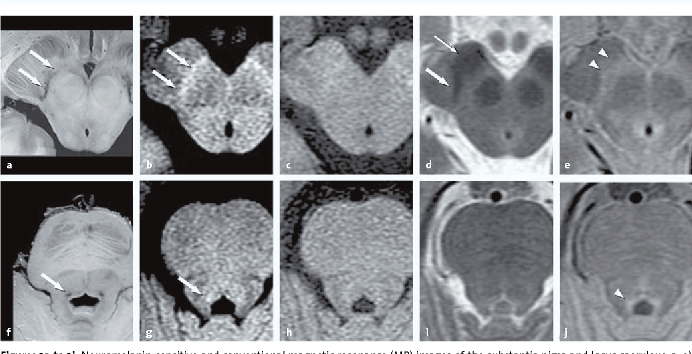
Figure 2 From Neuromelanin Sensitive Mri Semantic Scholar

Substantia Nigra Radiology Reference Article Radiopaedia Org

Substantia Nigra Anatomy On 3t Swi Mri Demonstrated Is A 3t Swi Download Scientific Diagram
Mri Of The Locus Coeruleus And Substantia Nigra Ppt Video Online Download
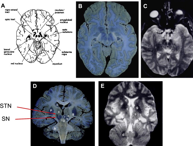
Anatomy Of The Substantia Nigra And Subthalamic Nucleus On Mr Imaging Radiology Key
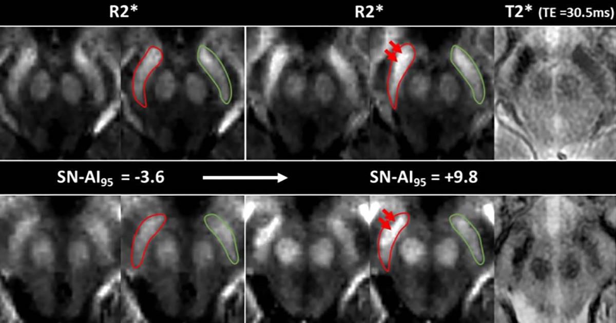
Auntminnie Com Called R2 Mapping The Mri Technique Can Detect Increased Iron Concentrations In The Substantia Nigra And Can Be Added To Follow Up Mri Scans To Monitor Neurodegeneration Radiology Rsna Radiology T Co Upjnjtcjq7

Comparative Study Of Mri Biomarkers In The Substantia Nigra To Discriminate Idiopathic Parkinson Disease American Journal Of Neuroradiology

Evaluation Of The Substantia Nigra In Patients With Parkinsonian Syndrome Accomplished Using Multishot Diffusion Weighted Mr Imaging American Journal Of Neuroradiology

Reproducibility Assessment Of Neuromelanin Sensitive Magnetic Resonance Imaging Protocols For Region Of Interest And Voxelwise Analyses Biorxiv
English T1 Weighted Mri Image Of A Brain Of A Patient Affected By Bpan Beta Propeller

Imaging Brain Structures That Deteriorate In Parkinson S Kurzweil
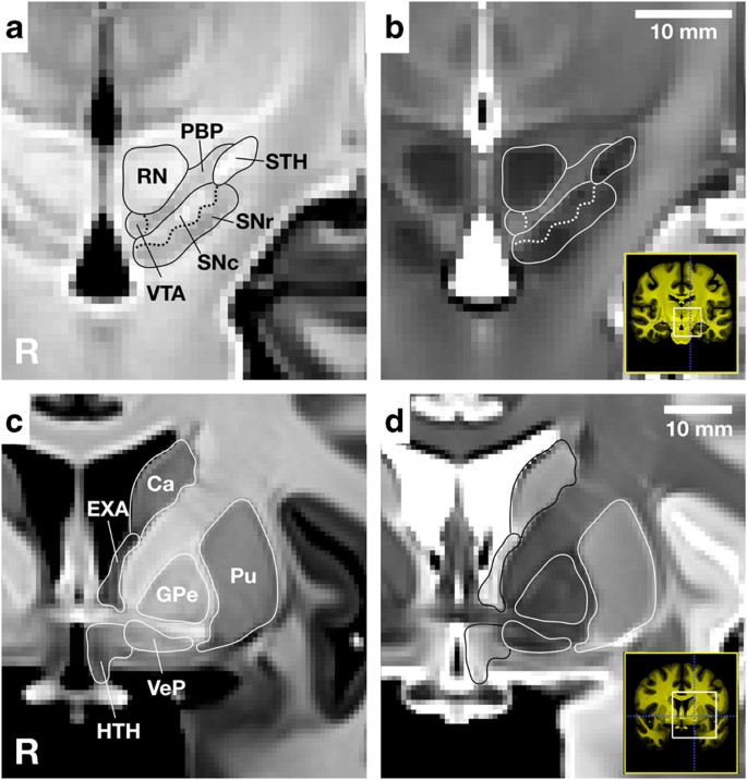
A High Resolution Probabilistic In Vivo Atlas Of Human Subcortical Brain Nuclei Scientific Data
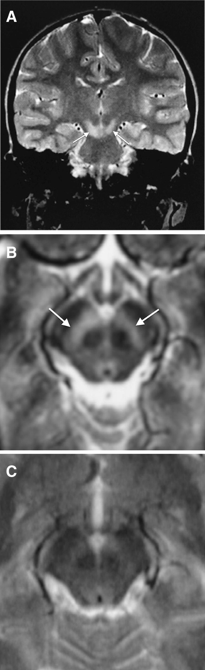
Isolated Substantia Nigra Lesions In Encephalitis A Specific Mri Pattern Springerlink
Plos One Nigrosome 1 Visibility At Susceptibility Weighted 7t Mri A Dependable Diagnostic Marker For Parkinson S Disease Or Merely An Inconsistent Age Dependent Imaging Finding

View Image

Evaluation Of The Substantia Nigra In Patients With Parkinsonian Syndrome Accomplished Using Multishot Diffusion Weighted Mr Imaging American Journal Of Neuroradiology
New Mri Tools Shed Light On Parkinson S Disease Progress Compact Mri System In Development For Improved Joint Imaging Diffusion Weighted Mri Could Guide Development Of New And Improved Tumor Models Cardiac Ct Reveals

St Louis Encephalitis Mri Wikidoc
Http Www Bioline Org Br Pdf Ni

Background Concepts Section 1 Rehabilitation In Movement Disorders

The Substantia Nigra In Parkinson Disease Proton Density Weighted Spin Echo And Fast Short Inversion Time Inversion Recovery Mr Findings American Journal Of Neuroradiology
Mri Findings Nbia
Imaging Degeneration Of The Substantia Nigra In Parkinson Disease With Inversion Recovery Mr Imaging American Journal Of Neuroradiology
Imaging And Behavior In Parkinson S Disease Structural Imaging Chapter 7 Neuropsychiatric And Cognitive Changes In Parkinson S Disease And Related Movement Disorders

Mri Axial T2 Weighted

Loss Of Substantia Nigra Hyperintensity On 7 Tesla Mri Of Parkinson S Disease Multiple System Atrophy And Progressive Supranuclear Palsy Sciencedirect

The Role Of High Field Magnetic Resonance Imaging In Parkinsonian Disorders Pushing The Boundaries Forward Lehericy 17 Movement Disorders Wiley Online Library

High Spatial Resolution Diffusion Mri In Parkinson Disease Lateral Asymmetry Of The Substantia Nigra Radiology

A B Axial T1 Weighted Magnetic Resonance Imaging Shows Symmetrical Download Scientific Diagram

Magnetic Resonance Imaging Of The Substantia Nigra In Parkinson S Disease Lehericy 12 Movement Disorders Wiley Online Library
Q Tbn And9gcrmsu3gc9tmucpxllu3db92ducrhhujnuxnr4 4p Oysr51me Usqp Cau
Diagnostic Imaging Of Bilateral Abnormalities Of The Basal Ganglia
Mri Of The Globus Pallidus And Substantia Nigra In Four Main Forms Of Download Scientific Diagram
Imaging Parkinsonian Pathology In Substantia Nigra With Mri Springerlink
Anatomical And Functional Organization Of The Human Substantia Nigra And Its Connections Elife

Detecting Dopaminergic Neuronal Degeneration Using Diffusion Tensor Imaging In A Rotenone Induced Rat Model Of Parkinson S Disease Fractional Anisotropy And Mean Diffusivity Values Liu Lx Du D Zheng T Fang Y Chen Ys
Q Tbn And9gcsgym3fllmvif Ded Bo1xh4okocoseq28gpsocnrhqky0mowdw Usqp Cau
Frontiers Combined Visualization Of Nigrosome 1 And Neuromelanin In The Substantia Nigra Using 3t Mri For The Differential Diagnosis Of Essential Tremor And De Novo Parkinson S Disease Neurology

Substantia Nigra Anatomy Radiology Case Radiopaedia Org

Bilateral Substantia Nigra Changes On Mri In A Patient With Encephalitis Lethargica Neurology

Iron Measurements With Mri Reveal Stroke S Impact On Brain Eurekalert Science News

Anatomy Of The Substantia Nigra And Subthalamic Nucleus On Mr Imaging Radiology Key

Reproducibility Assessment Of Neuromelanin Sensitive Magnetic Resonance Imaging Protocols For Region Of Interest And Voxelwise Analyses Biorxiv

A Mri T1 Weighted Image Shows Bilateral Hypointensities In Substantia Download Scientific Diagram

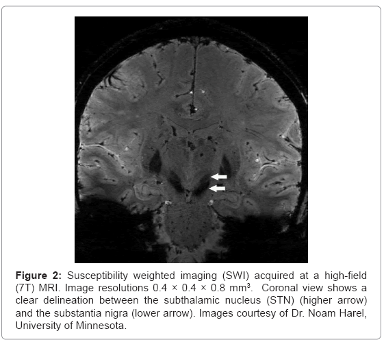
Susceptibility Weighted Imaging Swi Acquired At A Hig Open I

Xmlinkhub
Frontiers Chemical Exchange Saturation Transfer Mri Signal Loss Of The Substantia Nigra As An Imaging Biomarker To Evaluate The Diagnosis And Severity Of Parkinson S Disease Neuroscience

Mr Imaging Of The Substantia Nigra At 7 T Enables Diagnosis Of Parkinson Disease Radiology
Plos One Is R2 A New Mri Biomarker For The Progression Of Parkinson S Disease A Longitudinal Follow Up

Bilateral Hyperintense Lesions Of The Substantia Nigra On Mri Flair Download Scientific Diagram
Magnetic Resonance Imaging Mri In Parkinsons Disease Omics International

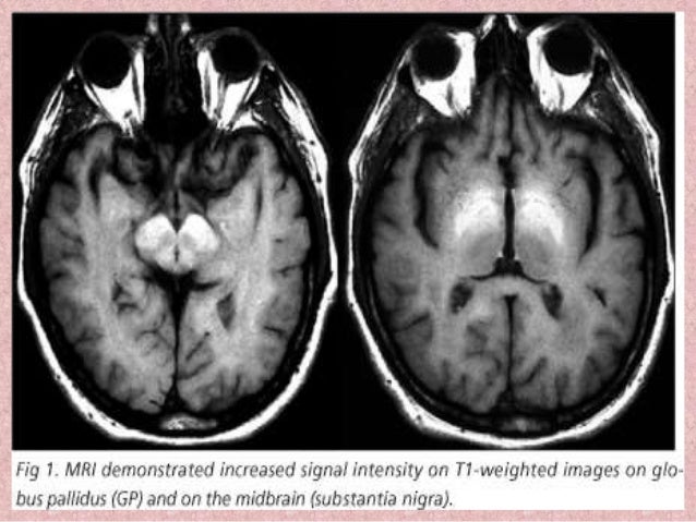
Chronic Manganese Toxicity Due To Substance Abuse In Turkish Patients

Substantia Nigra Wikipedia

The Substantia Nigra In Parkinson Disease Proton Density Weighted Spin Echo And Fast Short Inversion Time Inversion Recovery Mr Findings American Journal Of Neuroradiology

7 Tesla Magnetic Resonance Imaging A Closer Look At Substantia Nigra Anatomy In Parkinson S Disease Lehericy 14 Movement Disorders Wiley Online Library


